
All contents are concordant with mark schemes of CAIE 9700 Int’ A Level Biology past paper 4s from year 2014 to 2017 unless being noted with an asterisk (*).

This work is licensed under a Creative Commons Attribution-NonCommercial-NoDerivatives 4.0 International License.
本文作者/Authour(s):石天熠,肖博文
PDF Version: See here
Homeostasis in Humans in General
Define negative feedback
A process in which a change in some parameter, such as blood glucose level, brings about processes which move its level back towards normal again.
Outline how a negative feedback mechanism works
- A initial stimulus, which is the change in a parameter away from the set-point, is detected by receptors.
-
In response, hormones are released and they reach their target organs, or effectors.
- Corrective actions are taken by the effectors, allowing restoration of the ‘set point’.
Explain the principles of homeostasis in humans
Homeostasis is the maintenance of constant internal environment irrespective of changes in external environment, using negative feedback mechanisms. This involves the initial stimulus, which is the change in a parameter detected by receptors, and the action taken by the effectors, allowing restoration of the ‘set point’. There is fluctuation around the norm. Examples of homeostasis include thermoregulation, osmoregulation, and blood glucose regulation.
Examples and importance
Homeostasis is the maintenance of constant internal environment, for example several parameters of blood (e.g. blood glucose level, osmolarity and blood pressure), causing them to fluctuate within narrow ranges around the set point.
Homeostasis of body temperature, water potential (osmolarity) and blood glucose level is critical. Low body temperature leads to slowed metabolism and enzyme activity, while high temperature causes enzymes to be denatured; low water potential causes cells to shrink while high water potential causes cells to burst; low blood glucose level reduces cellular respiration while high blood glucose level leads to lowered blood water potential, which further leads to shrinkage of cells.
Human Kidney Anatomy and Physiology
Structure of A Kidney
- A kidney consists of the cortex on the outer side, the medulla and a pelvis. It is connected to the renal artery and the renal vein.
- Nephrons are the functional units of a kidney. A nephron consists of the renal capsule, the proximal convoluted tubule, the loop of Henle, the distal convoluted tubule and the collecting duct.
- The glomerulus is a network of capillaries surrounded by the renal capsule. The afferent arteriole enters it, and the efferent arteriole transports blood away from it.
Water Potential
Tendency of water molecules to move from one region to another
Ultrafiltration
The diameter of afferent arterioles is greater than that of the efferent arterioles, so this builds up a high blood pressure in the glomerulus and molecules tend to move from the capillary into the Bowman’s capsule.
Molecules first pass through holes in the endothelium, then the basement membrane, and finally gaps between podocytes and enter Bowman’s capsule. The basement membrane is selectively permeable: only molecules with molecular weight less than 69,000 can pass through it.
Features of Epithelial Cells of the Proximal Convoluted Tubule
- Microvilli and folded basal membrane: increase surface area for absorption of Na+/glucose/amino acids
- Many mitochondria: provide energy/ATP for Na+/K+ pumps
- Tight junction between cells: hold adjacent cells together; fluid cannot pass between cells/substance must pass through cells
- Many cotransporter proteins: for glucose reabsorption
- Many aquaporins: for water reabsorption
Mechanism of Reabsorption in the Proximal Convoluted Tubule
- Active transport using ATP by Na+/K+ pumps pump Na+ ions out of the cells into the blood. This sets up a concentration gradient of Na+.
- Facilitated diffusion using co-transporter proteins allows the transport of glucose, amino acids and ions from the lumen to the PCT cells against their concentration gradients, along with Na+ ions down its concentration gradient. In addition, osmosis occurs as water moves down a water potential gradient into the PCT cells.
Reabsorption in loop of Henle*
- Sodium and chloride ions are actively transported out of the cell in the upper (thicker) half of the ascending limb
- This lowers the water potential of the tissue fluid
- Water moves down the water potential gradient out of the loop of Henle in the descending loop
- The loss of water increases the concentration of ions in the loop
- Na+ and Cl+ ions diffuse out of the loop in the lower (thinner) part of the ascending limb down their concentration gradients
- This mechanism, known as a counter-current multiplier, enables the maximum concentration of solutes to be built up both inside and outside the tube at the bottom of the loop
Why an increase in the quantity of protein in the diet leads to an increase in the concentration of urea in the urine
1. Proteins are hydrolysed into amino acids in gut
2. Excess amino acids cannot be stored
3. Amino acids are deaminated in liver to produce urea
4. Urea in blood is filtered into nephrons by ultrafiltration
Production of ADH
The hypothalamus has neurosecretory cells, which produce ADH, and osmoreceptors, which detects changes in water potential of the blood.
Osmoreceptors shrink when water potential is low, and this causes ADH to be released from the posterior pituitary.
Role of ADH
- ADH decrease the volume of urine produced by affecting the collecting duct.
- Specifically, It increases the permeability to water of the collecting duct so more water move out of the lumen down the water potential gradient.
- It does so according to a series of molecular mechanisms:
- First, ADH binds to corresponding receptors on cell surface membranes
- Binding to receptors activates the second messenger system (using cAMP) which, in turn, activates a series of enzyme controlled reactions by means of phosphorylation. This amplifies the original signal.
- Finally, phosphorylated vesicles, which contains aquaporins, move to and fuse with the cell surface membrane, so that additional aquaporins are added to the cell surface membrane, making the membrane more permeable.
Process of deamination of amino acids and formation of urea*
Ammonium ions/ammonia is produced in the liver from excess amino acids, which is toxic to the body if they are allowed to accumulate. Ammonium ions/ammonia combine with carbon dioxide to form urea in urea (ornithine) cycle.
Hormonal Control of Female Reproductive System
Describe the role of hormones in the maintenance of the human menstrual cycle
- FSH and LH, which are released by the anterior pituitary gland, stimulates the development of Graafian follicle.
- The follicle produces oestrogen, whose concentration rises in the first 12 days of menstrual cycle, causing endometrium to thicken.
- Around day 14, the surge in LH and FSH concentration stimulates ovulation and LH stimulate development of corpus luteum.
- Corpus luteum produces progesterone, which causes further development of endometrium.
- If there is no fertilisation, the combined high concentration of progesterone and oestrogen inhibits release of LH and FSH, which is an example of negative feedback.
- Corpus luteum degenerates and concentration of progesterone falls, so endometrium break down and menstruation occurs.
Outline the role of progesterone in the human menstrual cycle
1. It is mostly secreted during the second half of the cycle (i.e. from day 14 onwards)
2. It maintains the thickness of the endometrium in preparation for implantation
4. inhibits GnRH production and hence development of new follicle
Describe the roles of LH in the menstrual cycle
1. It stimulates follicle to secrete oestrogen
3. Surge in LH secretion stimulates ovulation
5. It stimulates development of corpus luteum, which secretes progesterone
Role of oestrogen
1. Thickening of endometrium
2. Development of capillaries in the endometrium
3. Inhibition of FSH production
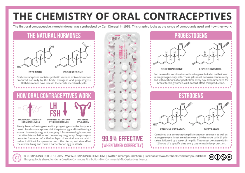
Pills
- Pills contain synthetic hormones that are equilavent to progesterone and oestrogen. Synthetic hormones are broken down more slowly so their activity last longer.
- Progesterone and oestrogen concontrations reamin high, and this reduce the secretion of FSH and LH from the anterior pituitary, which is a form of negative feedback. The lack of FSH prevents maturation of follicle, and the lack of LH prevents ovulation.
- Furthermore, more cervical mucus would be produced, which is hostile to the sperm, preventing implantation.
- Pills are taken daily for 21 days, then there is a 7-day break that allows menstruation before another 28-day cycle starts.
 Blood Glucose Level
Mechanism of Action of Adrenaline
- Adrenaline raises blood glucose level using a series of molecular mechanisms
- Adrenaline first binds to its receptors in cell surface membrane of target cells, for example liver cells
- The binding causes the receptor to change its conformation, which in turn activates G protein.
- G protein activates adenylyl cyclase, which produces cAMP.
- cAMP acts as a second messenger and activates kinases.
- These kinases activates other kinases by means of phosphorylation, and this forms an enzyme cascade, amplifying the original signal.
- Finally, a large amount of glycogen phosphorylase is activated, which breaks down glycogen into glucose, a process known as glycogenolysis.
- Glucose diffuses out of the cells into blood down the concentration gradient and via channel proteins, increasing blood glucose concentration
Response to High Blood Glucose Level
- High blood glucose level triggers a series of physiological reactions to lower blood glucose level back to the normal level, which is an example of negative feedback.
- High blood glucose level is detected by β cells in islets of Langerhans in pancreas.
- In response, these cells produce and secrete more insulin into blood, while production of glucagon in α cells is inhibited.
- Insulin increases glucose absorption in liver by phosphorylating glucose, and increases permeability to glucose in muscle cells by adding GLUT4 transporter proteins to their cell surface membranes.
- Insulin increases rate of respiration of glucose and conversion of glucose into glycogen.
Dip stick
- A dip stick is a pad containing immobilised glucose oxidase and peroxidase and chromogen, which is used to measure glucose concentration.
- A dip stick is lowered into the sample (blood, urine, etc.) and glucose oxidase oxidises glucose into gluconic acid (gluconolactone), producing hydrogen peroxide.
- hydrogen peroxide react with chromogen, a colourless compound (catalysed by peroxidase) to produce a brown colour. The darkness of the brown colour is proportional to the concentration of glucose.
Glucose Biosensor
- The biosensor contains a pad with immobilised glucose oxidase and peroxidase.
- The pad is lowered into the sample (blood, urine, etc.) and glucose oxidase oxidises glucose into gluconic acid (gluconolactone), producing hydrogen peroxide.
- Hydrogen peroxide is broken down by peroxidase, producing oxygen, which generates an electric current that can be detected by the electrodes to give a numerical value of glucose concentration.
Symptoms of Diabetes mellitus
- Feeling thirst
- Patients have high blood glucose concentration
- This causes decrease in water potential of blood
- Which is detected by osmoreceptors in the hypothalamus
- Loss of body mass
- Insulin function is compromised
- Less glucose is converted to fat and glycogen, and less glucose is taken up by cells for respiration
- More fats and proteins are respired
Homeostasis in Plants
Opening and Closing of Stomata
- Stomata open in response to:
- Increasing light intensity
- To gain CO2 for ph0tosynthesis
- To allow oxygen out
- Allows transpiration to occur, bringing water for photosynthesis
- Increasing light intensity
- Stomata close in response to:
- Darkness
- CO2 not required as no photosynthesis
- High temperature/low humidity/water stress
- Prevent water loss by transpiration
- Maintain cell turgidity/prevent wilting
- Darkness
Mechanism of Stomata Opening
- Stomata opening are dependent on the turgidity of guard cells, which involves a series of molecular mechanisms.
- There are proton pumps in cell surface membranes of guard cells that pump H+ out of the cells, lowering H+ concentration inside the cell. Therefore, the voltage inside the cell is more negative than the outside, building up a electrochemical gradient.
- K+ channels open and K+ move into the cell down the electrochemical gradient by facilitated diffusion. This is followed by entry of Cl– ions.
- Entry of K+ and Cl– ions lowers the water potential inside the cell, so water move into the cell via aquaporins by osmosis, down the water potential gradient.
- Volume of guard cells increases, and they become turgid. Guard cells have uneven thickness of cell walls so that turgidity would cause them to bent in a way that opens the stomata.
Role of Abscisic Acid (ABA)
Plants secrete abscisic acid at times of water stress. It closes the stomata to reduce water loss.
Abscisic acid binds to receptors on cell surface membrane of guard cells, inhibiting proton pumps. H+ concentration inside the cell increases.
Abscisic acid also stimulates the influx of Ca2+ ions, which act as a second messenger to activate channel protein to open, encouraging efflux of K+ ions.
Water potential of the cells increases, so water leaves cells by osmosis. Volume of the guard cells decrease and cells become flaccid.
Thermoregulation
Principles of Thermoregulation
- A change in blood temperature is detected by the hypothalamus.
- The brain sends impulses to effector, which carries out response (e.g. shivering; vasoconstriction/vasodilation),
- Actions taken by the effectors return blood temperature to normal.
- This is an example of negative feedback mechanism
Process of Thermoregulation
When temperature is low
- Vasoconstriction
- Arterioles in skin get narrower
- Less blood flow through surface capillaries
- So there is less heat loss to surroundings
- Shivering
- Involuntary muscle contraction
- Releases heat
- Increasing secretion of adrenaline
- Increases metabolic rate (i.e. rate of respiration)
- More heat released
When temperature is high
- Vasodilation
- Arterioles in skin gets wider
- More blood flow through surface capillaries
- So there is more heat loss to surroundings
- Sweating
- More sweat produced
- Sweat evaporates
- Using heat energy (latent heat of vaporisation)
